Osteo-articular diseases
The strength of the bone tissue is not simply related to bone density, but it also depends on the micro-architecture of the trabecular bone. Studies have shown that the structural and mechanical properties of spongy bone were anisotropic and have suggested using anisotropy to predict the risk of fracture [BILC+05]. Indeed, whatever the degree of accuracy of the characterization of a trabecular microarchitecture, the orientation of trabecular bone spans and the direction of mechanical tests are powerful features of the bone strength. On this basis, several researchs have been undertaken to model the trabecular microarchitecture from the observation of bone radiographs, and to characterize it using various anisotropy indices [CBIL+05]. The goal of such researchs is to predict the risk of fracture using one or several indices extracted from images or the combination of these indices with known clinical factors.
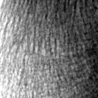
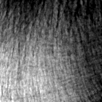
The analysis of the textures of bone radiographs is one of the approaches for the characterization of the trabecular micro-architecture. In the past, the fractal analysis, based on the model of isotropic fractional Brownian motion [BPL+01,BLJ+94] was used. In these works, the anisotropy is defined by a characterization of the shape of a so-called polar diagram [BLJ+94]. The analysis is used to compare a carrier bone (calcaneum) to a non-carrier bone (radius) [LJH+96]. Still, the interpretation of the rotational variations of texture irregularities are difficult and quite different from span directions. To remedy this defect, some authors have established a method for analyzing the anisotropy, called ``the method of directional averages'' in the framework of anisotropic fractional Brownian models [LHJ+04]. However, we could notice that the theoretical properties of these models were not observed on bone radiographs, suggesting that they models do not suit the images.
On the other hand, the bone texture anisotropy computed on projected images must be confronted to the anisotropy of the 3D object [PPL+00]. Our experiences have shown that alterations due to a random anisotropic structure increases its anisotropy [PLB01]. The investigation of such 2D-3D correspondence aspects is necessary for the validation of an anisotropy index, since the issue is to provide a relevant index of risk, which is related to 3D structures and takes into account the anisotropy. In particular, the trabecular bone is organized as a complex network of plates and beams, and recent studies [SM05] show that it is important to take into account these two components in order to get reliable indicators of the state of bone micro-architecture.
During these last years, we were concerned with the description of the 3D architecture of 3D human bones and the characterization of the topology and the morphology of numerous tomographic reconstructions [PPL+00,PBP+00,PLL+02,PRM+02]. We analyze the emergence and the evolution of the solid matrix anisotropy, as the initial trabecular structure progressively evolves according to some specific degradation scenarios [PLB01].