Breast cancer
The radiographic appearance of a breast depends primarily on the distribution of adipose and fibro-glandular tissues it contains. The mammographic density refers to the bright image aspect produced by the presence of fibro-glandular tissues. In the late 1960s, Dr. Wolfe suggested that the risk of developing a cancer was related to the radiographic appearance of breasts and their density [BOC+82,BMS+82,Wol67b,Wol67a]. In addition to the medical debate raised by this conjecture [HM02a,HM02b], the Wolfe's investigation has inspired a lot of works in Image Processing which aims at the automation of the quantification and the characterization of the density [BNBF+96,BYL+97,CSH+90].
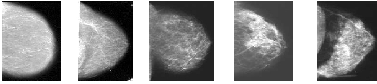
These works have taken several research directions including one which is based on the modeling of mammograms by fractional Brownian fields [BJJ01a,BNBF+96,CSH+90,HDV+99,HV02,KLSJ+01]. These models are particularly well adapted to mammograms. The Hurst index, which characterizes the regularity of these fields, is correlated with the mammographic density. However, these models are limited, due to the fact that they do not take into account the mammogram anisotropy. In a previous work [RB07], we showed that mammograms have an anisotropic nature and that anisotropic fractional Brownian fields are better suited for the modeling of mammograms than isotropic fractional Brownian fields.
In another perspective, some works have been conducted to assess the density of breasts from area measurements [BBF+94] and volume measurements [DWW+08,VESH+08]. High breast density is one of the strongest known risk factor for breast cancer. Women with over 75 percent of their breasts composed of dense tissues have an estimated 2-6 fold increase in the cancer risk, as compared to those with very low breast density. Several studies have also concluded to an increase in the mammographic density among women using menopause hormone therapies.
In this project, we focus on the mammogram aspect rather than the breast density itself. The mammogram aspect is characterized using parameters of a well-suited model of mammogram textures. We study whether such parameters provide an information which is complementary to the density measurements and investigate their association to a breast cancer risk using the database E3N.
Besides, the detectability of masses in a mammographic texture reveals surprising results, because at a given contrast level, the smallest masses are the most visible, contrarily to what happens in a white noise texture for example. This property of human vision [Kot98,BJJ01b,BJJ01a] is confirmed when mammographic textures are modeled by fractional Brownian fields and when an a-contrario detection model [DMM08] is used with almost any contextual measure. Such results may have consequences on the optimization of mammographic imaging, and on the improvement of computer-aided detection or vizualization tools.