MATAIM Research Project.
MATAIM is a multidisciplinary research program at junction of Image Processing, Probability Theory, Statistics, and Medecine. It focuses on Anisotropic Models of Textures and Applications to Medical Imaging.
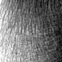
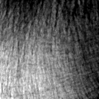
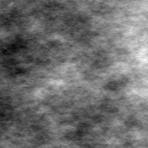
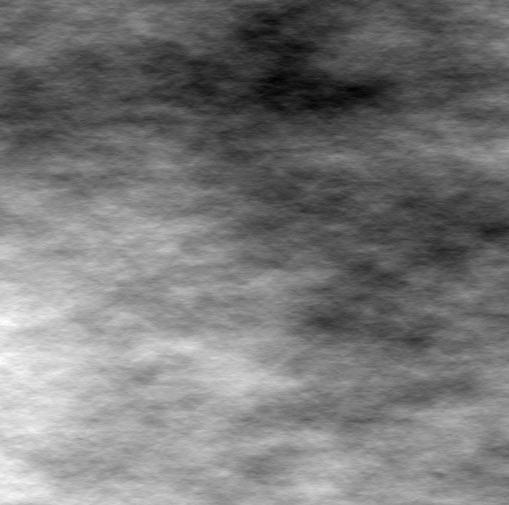
Texture analysis is one of the fundamental issues of Image Processing, which has many applications in medical imaging. In this project, texture analysis issues are tackled from a probabilistic view point, considering the image as a realization of a random field whose properties reflect those of the texture formation. Random fields models of textures contain some parameters which are used to characterize the textural aspect of images and extract some medical information from images. For instance, the fractal analysis , which was a great success in medical imaging [CDF89], has been applied either to mammography for the characterization of the breast density, the classification of breast types and the assessment of cancer risks [CSH+90,HM02b],or to bone radiographs for the characterization of the bone architecture and the evaluation of the osteoporotic fracture risk [BPL+01,BLJ+94].
In collaboration with our medical partners, we focus on two medical applications, which have a major importance to Public Health: the breast cancer and the osteo-articular diseases (osteoporosis, rheumatoid arthritis, inflammatory diseases,...).
The team INSERM U 658 (Regional Hospital Center of Orleans, France) is devoted to the characterization of bone tissue and the quality of bone architecture using imagery. Its main goal is to supplement the densitometry used in clinical practice for measuring the bone mass with a texture analysis of bone images which would give relevant information about the trabecular microarchitecture. A multicenter cross-national study of menopausal woman populations was conducted by [LGK+08] and a monitored prospective cohort is planned.
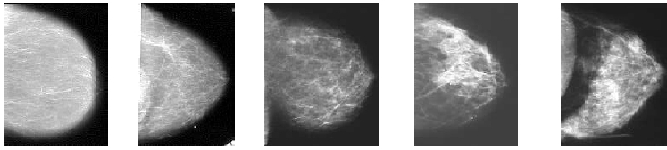
The unit INSERM ERI-20 (Gustave Roussy Institute in Villejuif, France) has an expertise in the epidemiology of the breast cancer. Its work is based on the survey data E3N, which results from a large cohort study involving 100 000 female volunteers. Using an updated database, the goal is to answer a range of challenging issues concerning the medical aspect of mammograms.
The previous applications raise a methodological problem which is at the forefront of our project: How to characterize relevantly the appearance of radiographs (mammograms or bone radiographs)? We use the analysis of texture to address this issue. The approach we follow consists in proposing random models for the modeling of radiological images and using the estimated parameters of models for the construction of indices which characterize the textural aspect of images. Due to the anisotropic nature of bone radiographs and mammograms, the modeling of these images is a difficult mathematical task.
|
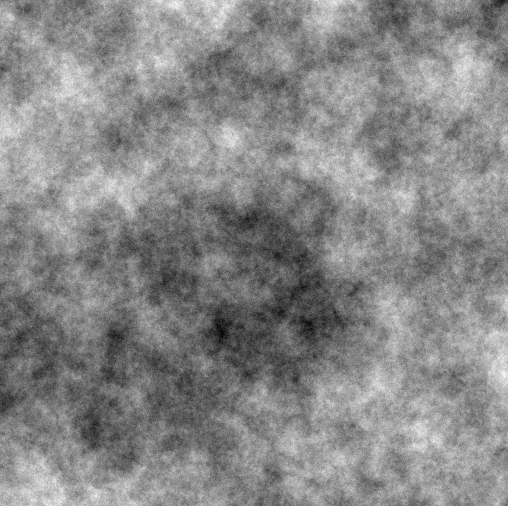

The theoretical study of anisotropic models raises numerous challenges which have to be taken up for succeeding in model applications. First, it is necessary to define mathematically the anisotropy and to know how to apprehend it through models. For many anisotropic models, it also remains to develop strategies for estimating model parameters and testing the adequacy of models to data. In addition, it still remains to design techniques for simulating accurately the defined random fields. Hence, the main part of the project is devoted to the mathematical study of these methodological issues. It involves researchers in Probability Theory and Image Processing mainly grouped into two mathematical teams : MAP5, CNRS UMR 8145 from University Paris Descartes and LTCI, CNRS UMR 5141 from the School of Telecommunications in Paris.
The research program is funded by the Agence Nationale pour la Recherche (ref. ANR-09-BLAN-0029-01), and the Institut National du Cancer (INCa) (ref. 2009-1-SHS SP-01-UP5-1).